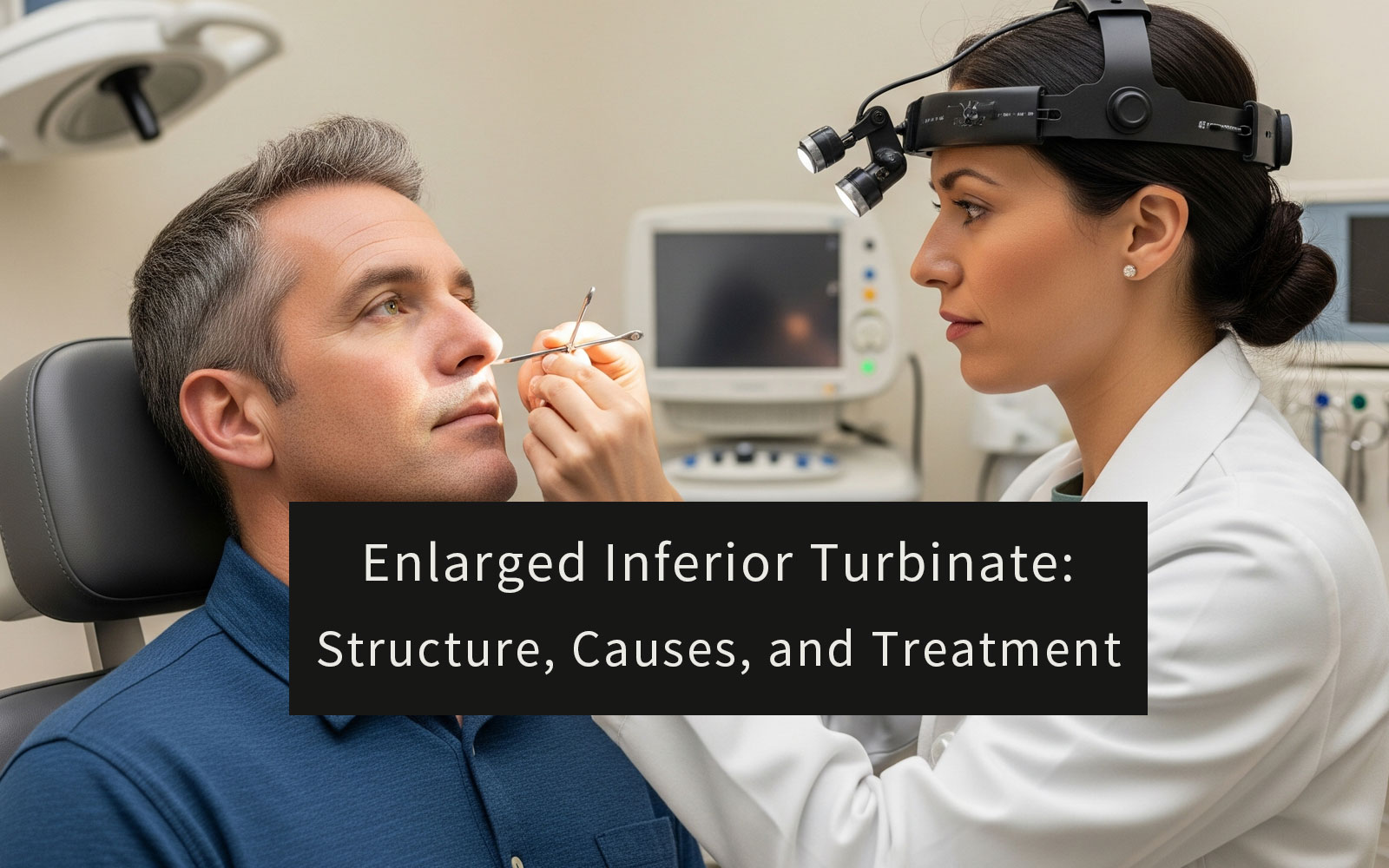
Enlarged inferior turbinates, also known as inferior turbinate hypertrophy, are a leading anatomical cause of chronic nasal congestion and nasal obstruction in both adults and children. Common symptoms include persistent nasal blockage and habitual mouth breathing, which may result from either structural abnormalities or ongoing inflammation of the inferior turbinates. Understanding the anatomy, primary causes of enlargement, and evidence-based treatment options is crucial—not only for ENT professionals but also for medical distributors seeking to improve patient outcomes and provide effective post-surgical care solutions. By recognizing and addressing inferior turbinate hypertrophy early, healthcare providers can significantly enhance patients’ breathing comfort and overall quality of life.
Table of Contents
What Is the Inferior Turbinate?
The nose contains bony structures called turbinates (also known as nasal conchae), each covered by a soft tissue layer called the nasal mucosa. There are three pairs of turbinates in the nasal cavity: inferior, middle, and superior. The inferior turbinate is the largest and most vital for healthy breathing. Located along the lower sidewall of each nasal passage, it extends horizontally and is a separate bone, unlike the middle and superior turbinates which are part of the ethmoid bone. The surface of the inferior turbinate is lined with highly vascular tissue, allowing it to swell or shrink naturally to regulate airflow—a process known as the nasal cycle. This dynamic function helps maintain optimal airflow and protects the respiratory tract.
Primary Functions of the Inferior Turbinate:
- Directing airflow: Helps channel air through the nasal passage
- Warming and humidifying: Adds heat and moisture to inhaled air
- Filtering: Traps dust, pollen, and particles through mucus secretion
Differences Among Inferior, Middle, and Superior Turbinates
While all turbinates share the role of conditioning inhaled air, they differ in structure, origin, and clinical significance. The inferior turbinate is the largest and most often involved in nasal obstruction. The middle turbinate helps protect sinus openings and supports olfactory functions, while the superior turbinate, being the smallest, mainly supports the olfactory bulb and is less involved in airflow. Understanding these differences is important for both diagnosis and treatment planning in ENT practice. The table below highlights the key differences:
| Feature | Inferior Turbinate | Middle Turbinate | Superior Turbinate |
|---|---|---|---|
| Location | Lowest, along the lateral nasal cavity wall | Above the inferior turbinate | Highest, above the middle turbinate |
| Bone Origin | Separate bone (inferior nasal concha bone) | Part of the ethmoid bone | Part of the ethmoid bone |
| Size | Largest | Intermediate | Smallest |
| Primary Role | Regulates nasal airflow; warms, humidifies, and filters air | Protects sinus openings; supports olfaction and airflow | Primarily supports the olfactory bulb |
| Associated Drainage | Inferior meatus – drains nasolacrimal duct | Middle meatus – drains maxillary, frontal, anterior ethmoid sinuses | Superior meatus – drains posterior ethmoid; sphenoid drains nearby |
| Clinical Significance | Most common cause of nasal obstruction when enlarged | Can contribute to blockage (e.g., concha bullosa, paradoxical curve) | Rarely obstructive; essential for olfaction |
| Additional Notes | Main target for turbinate reduction surgery | Key landmark in endoscopic sinus surgery | May be accompanied by a fourth (supreme) turbinate |
Symptoms of Enlarged Inferior Turbinates
Enlarged inferior turbinates, also referred to as turbinate hypertrophy, are most commonly associated with persistent nasal congestion and habitual mouth breathing. Patients may also experience a reduced sense of smell (hyposmia), frequent pressure-related headaches, and disrupted or poor-quality sleep. These symptoms occur because swollen inferior turbinates block normal airflow through the nasal passages, making it difficult to breathe comfortably through the nose.
Symptom Differences Across Age Groups
In children, enlarged inferior turbinates can interfere with feeding in infants and often lead to chronic mouth breathing. Over time, this may impact speech development, dental alignment, and facial growth. Sleep disturbances in children may also manifest as behavioral problems, irritability, or difficulty concentrating in school.
In adults, symptoms frequently result in chronic fatigue, reduced exercise tolerance, and a noticeable decline in overall quality of life. Ongoing nasal discomfort can negatively affect daily productivity, social interaction, and emotional well-being.
If you or your child experience these symptoms for an extended period, or if they become more severe, it is important to seek evaluation by an ear, nose, and throat (ENT) specialist. Prompt diagnosis and appropriate treatment can significantly improve nasal airflow and overall health.
How Enlarged or Inflamed Inferior Turbinates Lead to Nasal Congestion
The inferior turbinate is lined with highly vascular mucosal tissue that reacts quickly to environmental factors and immune triggers. When this tissue becomes enlarged or inflamed, commonly due to allergens, irritants, or compensatory changes related to nasal structure, the space inside the nasal passage narrows significantly. As a result, airflow is reduced and patients develop a sensation of constant nasal congestion or blockage.
This chronic narrowing of the airway can cause individuals to breathe through their mouths, especially during sleep. Mouth breathing may lead to symptoms such as dry mouth, sore throat, and an increased risk of sleep-related breathing disorders, including snoring or sleep apnea. Unlike nasal congestion from a common cold, which is typically temporary, turbinate hypertrophy tends to be long-lasting and may progressively worsen unless it is properly managed.
Recognizing the role of inferior turbinate hypertrophy in chronic nasal obstruction is essential for effective diagnosis and treatment. Timely medical or surgical intervention can restore normal airflow and help prevent long-term complications.
Essential Post-Op Care Checklist
| Care Step | Purpose | How to Do It / Tips |
|---|---|---|
| Nasal Irrigation | Flushes out mucus, blood, and crusts; maintains cleanliness | Start gentle saline rinses 24–48 hours after surgery (as instructed). Use sterile saline spray or premixed kits only. |
| Humidification & Moisturization | Prevents dryness and crusting, reduces irritation | Use a cool-mist humidifier and isotonic saline spray several times daily. Ask your doctor if you need nasal ointment. |
| Avoid Forceful Nose Blowing | Protects healing tissue and nasal packing | Gently sniff if needed. Sneeze with your mouth open to reduce pressure. Never blow your nose forcefully. |
| Limit Strenuous Activity | Reduces risk of bleeding or swelling | Avoid heavy lifting, bending, or vigorous exercise for at least 1–2 weeks. Consult your doctor for clearance. |
| Attend Follow-Up Visits | Ensures safe healing and professional care | Go to all scheduled appointments. Never remove or adjust nasal packing on your own. |
| Ask About Absorbable Dressings | Enhances comfort and speeds up recovery | Products like NasoAid® dissolve naturally, reduce discomfort, and support moist healing. |
The Relation Between Inflammation and Enlarged Inferior Turbinates
Inflammation is one of the most common causes of inferior turbinate swelling. Chronic conditions such as allergic rhinitis can trigger ongoing inflammation and increased blood flow in the nasal mucosa, leading to persistent turbinate hypertrophy. However, not all cases of enlarged inferior turbinates are purely inflammatory. In some instances, long-term changes to the underlying bone structure may occur, resulting in permanent enlargement that does not resolve even after inflammation is controlled.
From a clinical perspective, it is essential to distinguish between reversible mucosal hypertrophy and fixed anatomical enlargement. This distinction guides treatment decisions. While reversible cases may respond well to medications like intranasal corticosteroids, fixed or structural hypertrophy may require surgical intervention for lasting relief. Accurate assessment helps ensure patients receive the most appropriate and effective care for their nasal obstruction.
Common Causes of Enlarged Inferior Turbinates
Enlarged inferior turbinates, or inferior turbinate hypertrophy, can result from a variety of underlying causes. Identifying the root cause is essential for selecting the most effective treatment. The most common factors include:
1. Chronic Allergic Rhinitis
Long-term exposure to allergens such as pollen, pet dander, or dust mites can cause ongoing inflammation of the nasal mucosa. In allergic individuals, the immune system overreacts to these substances, increasing blood flow and mucus production within the turbinates. Over time, this repeated inflammatory response results in swollen turbinates and chronic nasal congestion. Seasonal variation is common, though some patients experience year-round symptoms depending on exposure patterns.
2. Compensatory Hypertrophy from Septal Deviation
When the nasal septum is misaligned or deviated, it reduces airflow on one side of the nasal cavity. To compensate, the inferior turbinate on the opposite side may enlarge in an attempt to equalize airflow resistance. This compensatory turbinate hypertrophy is structural in nature and often unresponsive to medication. It frequently persists unless both the turbinate and the deviated septum are surgically addressed.
3. Chronic Inflammatory Rhinitis
Some individuals experience turbinate swelling without any identifiable allergic cause. This condition is referred to as non-allergic rhinitis or vasomotor rhinitis. It is thought to result from abnormal nerve activity that affects blood vessel tone in the nasal mucosa. Common triggers include cold air, changes in humidity, strong odors, or hormonal fluctuations. Swelling in this case may come and go throughout the day and can lead to a persistent feeling of nasal congestion.ction.
4. Environmental Irritants
Exposure to air pollution, cigarette smoke, workplace chemicals, or other airborne toxins can irritate the nasal lining, promoting long-term inflammation and turbinate hypertrophy. Urban and industrial environments may increase this risk.
5. Repetitive Airflow Trauma
When nasal airflow is altered—due to septal deviation or narrowed passages—the air may strike the turbinate surface unevenly. Over time, this localized friction can stimulate the tissue to thicken and harden, much like how skin develops a callus under pressure. This mechanical stimulation contributes to a form of turbinate hypertrophy that is structural and non-reversible with medication alone.
6. Chronic Sinus Infections
Recurrent or untreated rhinosinusitis can lead to adjacent inflammation of the nasal turbinates. Infections that persist in the paranasal sinuses may cause mucosal edema in surrounding nasal structures, especially the inferior turbinate, which is closest to the sinus drainage pathways. In chronic cases, inflammation may lead to tissue remodeling and long-term enlargement.
7. Bony Remodeling and Structural Changes
In some cases, turbinate hypertrophy involves not only soft tissue swelling but also changes to the underlying bony structure. These changes may develop over time due to developmental abnormalities or prolonged inflammation. Once the turbinate bone thickens, the hypertrophy becomes fixed and typically requires surgical intervention such as turbinate reduction.
Not all enlarged inferior turbinates are caused by inflammation. Some are reactive and reversible, while others involve permanent anatomical changes. Differentiating between these causes is critical for selecting appropriate treatment, whether through medications like intranasal corticosteroids or procedural options such as radiofrequency reduction or submucosal resection. Accurate diagnosis not only improves patient outcomes but also reduces the risk of unnecessary or ineffective interventions.
Treatment for Enlarged Inferior Turbinates
Non-Surgical Treatment Options
Non-surgical therapy is most effective for cases driven by inflammation, allergy, or environmental factors. These approaches aim to reduce turbinate swelling while preserving nasal structure.
Intranasal Corticosteroids
Topical nasal steroids help reduce inflammation and mucosal swelling. They are commonly prescribed for allergic rhinitis or chronic rhinosinusitis. With consistent use, symptoms often improve within a few weeks.
Antihistamines and Decongestants
Oral or intranasal antihistamines manage allergic reactions that lead to swollen turbinates. Short-term decongestants may also offer temporary relief by constricting blood vessels in the nasal mucosa. However, prolonged use of decongestants can cause rebound congestion.
Saline Nasal Irrigation
Regular rinsing with saline solution clears allergens and irritants, keeps the mucosal surface moist, and supports nasal hygiene. It is a safe adjunctive therapy suitable for all age groups.
These treatments are often used in combination and monitored over time to determine their effectiveness. When improvement is insufficient or symptoms recur frequently, surgical solutions are considered.
Surgical Treatment Options
Surgical intervention is recommended for patients with moderate to severe turbinate hypertrophy that has not improved with medication. Procedures vary in complexity and are selected based on anatomical findings and symptom severity. Please refer to the Turbinate Reduction Surgery section for detailed descriptions.
Minimally Invasive Techniques
Minimally invasive surgeries are ideal for mild to moderate cases and are associated with shorter recovery times. They include Radiofrequency Ablation (RFA), Coblation, Cryoturbinectomy, Cauterization, and Laser Turbinate Reduction. These techniques shrink turbinate tissue while preserving the mucosa, helping improve nasal airflow with minimal trauma.
Submucosal Resection (Microdebrider Turbinoplasty)
This procedure removes part of the turbinate bone and soft tissue beneath the mucosa using a powered microdebrider. It is typically used for patients with structural hypertrophy. The mucosal lining remains intact, which reduces complications and supports faster healing.
Partial Inferior Turbinectomy
Partial resection removes a section of the turbinate including both soft tissue and bone. It offers greater airway relief but carries a higher chance of nasal dryness or crusting. This option is generally reserved for patients with long-standing, severe obstruction.
Total Inferior Turbinectomy
This is a more radical procedure that removes most or all of the inferior turbinate. Although it can maximize nasal airflow, the risk of complications such as chronic dryness or Empty Nose Syndrome (ENS) is higher. As a result, it is performed only in select cases where all other treatments have failed.
Outfracture Technique
Outfracture involves gently repositioning the turbinate outward to create more space in the nasal airway. It is often combined with other procedures and is a low-risk option that can enhance surgical results when used appropriately.
Comparison of Inferior Turbinate Treatment Methods
| Treatment Option | Indication | Advantages | Disadvantages | Recovery Time |
|---|---|---|---|---|
| Intranasal corticosteroids | Inflammation, allergy-related hypertrophy | Non-invasive, effective, minimal side effects | Requires consistent long-term use, not effective for everyone | None |
| Antihistamines / Decongestants | Allergy, temporary nasal congestion | Convenient, quick symptom relief | Decongestants not for long-term use, rebound congestion possible | None |
| Saline nasal irrigation | Suitable for all ages | Safe, very low side effects, can be combined with other therapies | Does not cure underlying cause, requires ongoing use | None |
| Minimally invasive procedures (RFA, Coblation, Cryoturbinectomy, Laser) |
Mild to moderate structural hypertrophy | Minimal trauma, quick recovery, preserves mucosa | May require repeat treatments, variable effectiveness | 1–7 days |
| Submucosal resection (Microdebrider turbinoplasty) |
Structural hypertrophy | Preserves mucosa, durable results, faster healing | Requires anesthesia, some risk of bleeding/infection | 7–14 days |
| Partial inferior turbinectomy | Long-standing, severe obstruction | Significant improvement in airflow | Increased risk of dryness, crusting | 1–2 weeks |
| Total inferior turbinectomy | Severe cases unresponsive to other treatments | Maximizes airflow, resolves most hypertrophy | High risk of dryness, possible Empty Nose Syndrome | Over 2 weeks |
| Outfracture technique | Adjunct to other procedures | Very low trauma, low risk | Limited effect if used alone | 1–3 days |
These treatment options should be selected based on individual patient needs and discussed thoroughly with an ENT specialist. Regular follow-up and postoperative care are essential to optimize outcomes and minimize complications.
How Physicians Select the Right Treatment
Physicians evaluate each patient individually to determine the most appropriate treatment strategy. Key considerations include the duration and severity of nasal obstruction, the degree of turbinate hypertrophy (soft tissue versus bony overgrowth), coexisting conditions such as a deviated septum or allergic rhinitis, and previous treatment response. Imaging studies and nasal endoscopy provide valuable insights into structural factors. Patient preference, age, general health, and tolerance for surgery also influence treatment decisions. The goal is to restore nasal airflow while minimizing risks and preserving the natural function of the turbinates.
Learn more about turbinate reduction surgery
Nasal Dressing Supports Post-Surgery Recovery After Turbinate Reduction
Following turbinate reduction surgery, effective post-operative care is essential to ensure proper healing and reduce complications such as bleeding, crusting, or mucosal adhesions. Among the various post-surgical considerations, the use of an absorbable nasal dressing plays a particularly important role. Compared to traditional non-resorbable packing, absorbable materials can offer better hemostasis, maintain a moist healing environment, and significantly enhance patient comfort by eliminating the need for painful removal.
NasoAid® is a resorbable nasal dressing specifically designed for such applications. Composed of 70% collagen and 30% carboxymethyl cellulose (CMC), it provides excellent hemostatic performance while forming a protective barrier over the healing mucosa. The collagen supports natural tissue regeneration, while CMC promotes moisture retention and reduces crust formation. As NasoAid® gradually dissolves in the nasal cavity, it avoids the discomfort associated with traditional packing and supports a smoother, more comfortable recovery process. Its biocompatibility and ease of use make it a preferred option for ENT surgeons across both outpatient clinics and surgical centers.
